Light blogging: Untangling the white matter June 8, 2007
Posted by Johan in Neuroscience.trackback
While functional MRI gets most of the spotlight, another interesting MRI technique is diffusion tensor imaging, which allows you to look at the direction of the axons that constitute the white matter. I won’t embarrass myself by trying to explain the physics behind it, but essentially it works by tracking the movement of water molecules, which are known to move preferentially down the length of the axon, thus telling you which way the axon is going. I hasten to add that because of the limitations of current scanners, there is no way that you can look at individual axons this way – what you get is a kind of average of the directionality in each voxel (ie, a little cube of the brain).
I found a very nice online demonstration of brain connectivity that was created with diffusion tensor imaging. It plays right in your web browser, but be patient as it takes time to load.
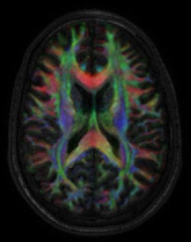
The colours you get are actually representations of the directionality: red stands for left-right, blue for superior-inferior (ie top-down), green for anterior-posterior (front-back). It’s a good idea to choose a second view by using the “Caption” drop-down menu on the right, and to untick the “link slices” box, so that you can move each view independently of the other, enabling you to move in 3 dimensions.
As you might expect, the picture that emerges isn’t exactly crystal clear. Some parts appear rainbow-like, suggesting that the wiring is going in all kinds of directions. A good example of something fairly straight forward is to go up in the axial view for a bit, until you see a huge red (ie, left-right) shape in the centre of the brain. This is the corpus callosum, which connects the two hemispheres.
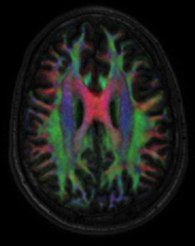
Or that’s the textbook version, at least. Go down instead for a bit, and note that some more red connections appear in more inferior locations – you get a good view of one in the axial slice that shows the eyes as complete globes. Clearly, there are a number of other crossover points between the two hemispheres in these subcortical areas.

It’s worth noting that the so-called “split brain” patients generally only had the corpus callosum severed (and sometimes this disconnection was not complete either), leaving these more inferior commissures intact. Despite the considerable deficits that these participants exhibited, parts of their brains actually remained connected, presumably enabling the surprising degree of every-day functioning that they actually had.
Update, June 10 2007:
I mixed up the URL to the DTI atlas. It should work now (thanks Miss Puzzlebrain!)

Comments»
No comments yet — be the first.